YAMOUT OPTICAL CENTER
|
YAMOUT OPTICAL CENTER |
|
Product
Optical historical |
Glaucoma Glaucoma is a progressive eye condition characterized by abnormally high intra-ocular pressure that can ultimately result in optic nerve damage. Glaucoma takes on many forms. Some types of glaucoma are secondary to associative systemic disease, while other types appear following surgery or physical injury to the eye. Certain medications may also induce glaucoma. The greatest risk potential for developing glaucoma, however, is a family history of the disease. Here is a general list of high-risk factors... · Family
history of glaucoma *Certain
mydriatic drugs and herbs, steroids, antihistamines, Understanding Glaucoma - The Anterior Chamber In order to understand how abnormal eye pressure develops, let's review the structures of the eye that are associated with fluid production and control.
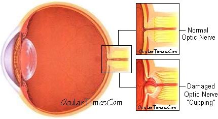
Within the eye is a mechanism for the continuous production and drainage of fluid. This fluid, called aqueous humor, is produced by a tiny gland, called the ciliary body, situated behind the iris. This aqueous fluid flows between the iris and lens into the anterior chamber, where it nourishes the lens and cornea, then flows out through tiny drains called the trabecular meshwork, and onward to the venous system. All eyes have internal pressure. The amount of pressure inside the eye is the balance between this fluid production and fluid drainage to and from the eye. Decreased fluid flow through the trabecular meshwork results in backup of fluid and elevated pressure within the eye. The Optic Nerve The optic nerve is the part of the eye that carries visual information to the brain. It is made up of over one million nerve cells, and while each cell is several inches long, it is extremely thin -- about one twenty-thousandth of an inch in diameter. When the pressure in the eye builds, the nerve cells become compressed, damaged, and ultimately destroyed. The death of these cells results in permanent visual loss. Relationship Of Intraocular Pressure (IOP) And The Optic Nerve Intraocular pressure is exerted on all walls of the eye, including the optic nerve and its blood vessels. The optic nerve is supplied with blood branches extending from the ophthalmic artery. If pressure in the eye is too high, blood may be prevented from adequately perfusing the optic nerve. If prolonged, this blood deficiency can permanently damage the optic nerve.
The typical optic nerve damage that occurs in glaucoma is known as "cupping." As the cells making up the nerve die, due at least in part to a pressure inside the eye that is too great for that particular eye to tolerate, they die and disappear. When sufficient numbers of these cells are gone, they leave behind a small crater or "cup" in the nerve. A portion of the nerve then appears to have been "scooped out." So one important thing doctors look for when they examine the optic nerve is the presence and extent of the "cup," how deep and wide it is. The Two Major Forms of Glaucoma ·
Open-angle
Glaucoma The Glaucoma Examination - Testing Methods A typical test method used to measure IOP is called applanation tonometry. The tonometer measures the amount of force needed to indent a small, central portion of the cornea. The test is painless, and takes only a few seconds to complete.
The average intraocular pressure (IOP) is about 14-16 millimeters of mercury (mmHg), but up to 21mmHg is regarded as within the normal range for most patients. A pressure of 22mmHg is defined as the borderline reading for a "glaucoma suspect". It is important to note that not everyone with a high IOP will necessarily develop visual damage or require treatment. In some cases, however, patients with normal IOP readings may develop a form of glaucoma known as "normal-tension" glaucoma. Thus, the diagnosis of glaucoma is not always determined by pressure readings alone. Damage to the
optic nerve results in visual field loss. Such loss can
become severe over time. Detection of glaucomatous visual
field loss is accomplished by visual field testing. See also retinal laser tomography for additional diagnostic glaucoma testing information. |
|
|

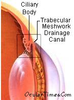 There are three fluid cavities in the eye:
the vitreous chamber, the posterior chamber,
and the anterior chamber. The anterior chamber
is situated between the cornea and the lens. It is
important to review the parts of the anterior chamber and
their relationship to one another in order to understand
the causes of glaucoma and its treatment.
There are three fluid cavities in the eye:
the vitreous chamber, the posterior chamber,
and the anterior chamber. The anterior chamber
is situated between the cornea and the lens. It is
important to review the parts of the anterior chamber and
their relationship to one another in order to understand
the causes of glaucoma and its treatment.