YAMOUT OPTICAL CENTER
|
YAMOUT OPTICAL CENTER |
|
Product
Optical historical |
Corneal Topography
Corneal topography maps graphically present a global picture of the corneal curvature. Most CT maps offer a two-dimensional representation of a three-dimensional shape. Colors are used to represent curvature values, and from these various color-coded curvatures, corneal shapes are categorized. Cool colors (blues) represent flatter curvatures and hot colors (reds) depict steeper curvatures. Interpretation of Map Images The color-coded display generated by the computer is a dioptric map, with each color representing a specific dioptric value relative to the corneal radius of curvature. With some systems, you can adjust the dioptric gradations from one color to the next. Warm colors usually represent higher amounts of curvature (steeper values), while cool colors represent flatter curvatures. Basic
Interpretation of corneal topographical maps are...
Astigmatic Curvature:
Most maps fall into five basic shape categories: round, oval, symmetric bow tie, asymmetric bow tie and irregular. The regular corneal surface is really an asymmetric asphere, like an off-center ellipse with toricity incorporated. The normal cornea typically results in a map with an aspheric and asymmetric topography. Some instruments refer to or model the cornea as a spline curve. Classically, the cornea is steepest centrally and flattens toward the periphery. Maps of both eyes of a normal individual, although similar, will not necessarily be mirror images of one another. There can be variations in normal corneal topography, but they are typically round or oval.
Normal
corneas and corneas affected by rigid contact lens wear
can produce a topographic pattern similar to that of
keratoconic eyes where the superior cornea is flatter
than the inferior cornea. In the absence of slit lamp
signs, these maps often lead to a diagnosis of
keratoconus suspect or pre-clinical keratoconus. Monitor
these eyes for changes. Mandell has shown that the apex
of the cone may be much steeper than a sagittal map
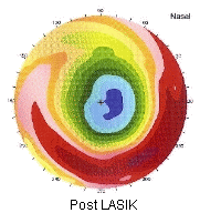
indicates. Refractive surgeries such as photorefractive keratectomy, radial keratotomy and LASIK result in topographical patterns that show central flattening and steepening toward the periphery, the inverse of the typical pattern of a normal cornea. Central islands resulting from such surgeries may not be detectable with keratometry, but are easily detectable by topography. Many topography systems also produce an astigmatic profile graph, a data overview display, and a keratometric display. This topographical analysis information can be used to track corneal healing following refractive surgery. Other Features Most topographers now offer a contact lens fitting analysis software program that provides a simulated fluorescein stain pattern to aid practioners in designing a rigid contact lens for patients. These systems also assess apical clearance and tear exchange between the cornea and posterior lens surface, allowing for a more accurate and accommodating lens fit that decreases the potential risks for contact lens related ocular complications. |
|
|


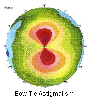 Astigmatism can be congenital
(with-the-rule, against-the-rule or oblique), contact
lens induced, or surgically induced. Regular astigmatism
is generally represented by a figure-eight or bow tie
pattern of increased corneal power that is vertical for
with-the-rule astigmatism and horizontal for
against-the-rule astigmatism. The two halves of the bow
tie are usually equal in size and 180 degrees apart.
Cataract surgery, penetrating keratoplasty and
trabeculectomy can also result in asymmetric oblique bow
tie patterns or irregular corneal maps. Trauma can also
cause a large amount of corneal irregularity. Tear film
abnormalities and corneal epithelial disruption can also
be detected by evaluating the videokeratography used in
most topography instruments. Conditions such as
keratoconjunctivitis sicca, infectious keratitis, toxic
epitheliopathies, epithelial basement membrane dystrophy,
Reis-Buckler's dystrophy and lattice dystrophy can also
show irregular patterns that are accompanied by decreased
visual acuity.
Astigmatism can be congenital
(with-the-rule, against-the-rule or oblique), contact
lens induced, or surgically induced. Regular astigmatism
is generally represented by a figure-eight or bow tie
pattern of increased corneal power that is vertical for
with-the-rule astigmatism and horizontal for
against-the-rule astigmatism. The two halves of the bow
tie are usually equal in size and 180 degrees apart.
Cataract surgery, penetrating keratoplasty and
trabeculectomy can also result in asymmetric oblique bow
tie patterns or irregular corneal maps. Trauma can also
cause a large amount of corneal irregularity. Tear film
abnormalities and corneal epithelial disruption can also
be detected by evaluating the videokeratography used in
most topography instruments. Conditions such as
keratoconjunctivitis sicca, infectious keratitis, toxic
epitheliopathies, epithelial basement membrane dystrophy,
Reis-Buckler's dystrophy and lattice dystrophy can also
show irregular patterns that are accompanied by decreased
visual acuity.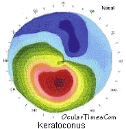 Keratoconus
can result in extremely complex and variable
topographical maps, most typically showing areas of
inferior steepening. The cone can assume various shapes
and sizes, and the apex can be at various locations in
relation to the central cornea. Although you cannot
diagnose keratoconus with topography alone, a
topographical map in combination with slit lamp signs
such as a Fleischer's ring, striae and/or corneal
scarring, will aid in diagnosis.
Keratoconus
can result in extremely complex and variable
topographical maps, most typically showing areas of
inferior steepening. The cone can assume various shapes
and sizes, and the apex can be at various locations in
relation to the central cornea. Although you cannot
diagnose keratoconus with topography alone, a
topographical map in combination with slit lamp signs
such as a Fleischer's ring, striae and/or corneal
scarring, will aid in diagnosis.