YAMOUT OPTICAL CENTER
|
YAMOUT OPTICAL CENTER |
|
Product
Optical historical |
Visual Field Testing Other Testing Methods Visual field testing may be kinetic or static. In kinetic testing (i.e., Goldmann or Tangent Screen), the stimulus is moved to different areas and the point at which it is first seen by the patient is marked. In static (stationary) perimetry, a specific point is chosen for examination and the stimulus is increased until its threshold is determined. Moving Targets Targets are moved from where you can't see them (beyond your side·vision) in towards the center of vision until you do see them. The test can be done with either a black screen on a wall (Tangent testing) or with a large bowl-shaped instrument (Goldmann testing). *Tangent
Screen Testing is rarely used today. Goldmann
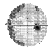
Visual Field With Goldmann or "kinetic" perimetry, a trained perimetrist moves the stimulus; stimulus brightness is held constant. The limits of the visual field are mapped to lights of different sizes and brightness. Important Measuring Tool These maps of visual sensitivity, made by either of fixed or moving targets, are very important in diagnosing diseases of the visual system. Different diseases of the eye and optic nerve show characteristic patterns of visual loss. The progression of glaucoma can be seen in these two examples. The darker areas on the visual field map suggest areas of progressive vision loss.
The visual field test is one of several important tools used to manage glaucoma. Certain diseases will damage the visual field. Glaucoma is one of those diseases. Visual field testing can detect glaucoma damage even before the patient is aware of it. The earlier glaucoma is detected, the sooner treatment can be started to stop damage.
There are many reasons other than glaucoma for an abnormal visual field result: the test was poorly given, the instrument was defective, the patient did not understand how to take the test, the patient was tired, the defect was real but does not indicate pathology, the defect is accounted for by some pathology other than glaucoma, eg, brain tumor, multiple sclerosis, a vascular problem, a congenital defect, an infection, or retinal disease such as macular degeneration, retinal detachment, or inflammation. Or the defect could be a false defect, that is, really not present at all! Despite all of the shortcomings of visual field testing it is the only way to document actual visual loss and whether such loss is progressing or remaining stable. As such it plays an indispensable role in helping glaucoma patients retain their sight. How Often is Visual Field Testing Performed? Visual field testing may be done every few months to every few years. The frequency of testing depends on the severity of retinal damage and whether or not the ocular disease is well-controlled. |
|
|